hpxmri@gmail.com - contact@pulmonaryimaging.com
TR +90 531 741 03 94
NEWS
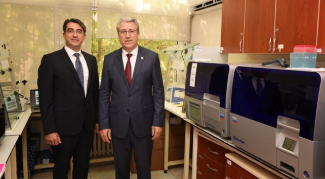
Ozkan Doganay, who left his career at Oxford University and worked on lung imaging devices at Ege University, where he graduated, is leading a young research group of 20 people. Doganay, 38 and father of two from Aegean Izmir province, graduated from Ege University Physics Department in 2007 and received his master's degree from Ryerson University in Toronto, Canada, with a scholarship. Working on lung imaging systems, Doganay worked as a research assistant at the same university for a year after his master's degree. The Turkish scientist completed his doctorate at the University of Western Ontario and won a national cancer research scholarship with his studies in Canada...
Ege Üniversitesinde, Dr. Özkan Doğanay, yüksek lisans öğrencisi Nur Ekenel ile Tıp Fakültesi öğrencileri Belkıs Aysu Özbek ve Ceren Yürümez, COVID-19'un akciğerlere etkisini saptamak üzere "COVID-19'da Bilgisayarlı Tomografi Yazılımlarının Geliştirilmesi" projesini tamamladı. Dr. Özkan Doğanay, TÜBİTAK desteği de alan proje kapsamında geliştirilen yazılımın hastaya tanı koymada, hastalığın hangi evrede olduğunu saptamada etkili olduğunu anlattı. Doğanay, "Yazılım, hastalığın akciğerde yayılışının üç boyutlu olarak görülmesine olanak sağlayacak ve hastalığın ciddiyeti ve gelişimi izlenebilecek. Bu tür yazılımlar Kronik Obstrüktif Akciğer Hastalığı (KOAH) gibi heterojen akciğer hastalıklarında ve akciğer kanserinde de kullanılabilecek" dedi.

Respiratory diseases such as lung cancer, asthma and chronic obstructive pulmonary disease (COPD) are a leading cause of death and disability in the world*. Early detection is essential — and one of Ryerson’s own is at the forefront of novel imaging techniques to that end. Since his days at Ryerson, Ozkan Doganay (MSc, Biomedical Physics ’10) has enjoyed success that has taken him around the globe — from Canada, to Oxford University in the U.K., and back home to Turkey, where he’s now building the country’s caliber in scientific research and innovation. ...

PUBLICATIONS
Ozkan Doganay, Trevor Wade, Elaine Hegarty, Charles McKenzie, Rolf F. Schulte, and Giles E. Santyr
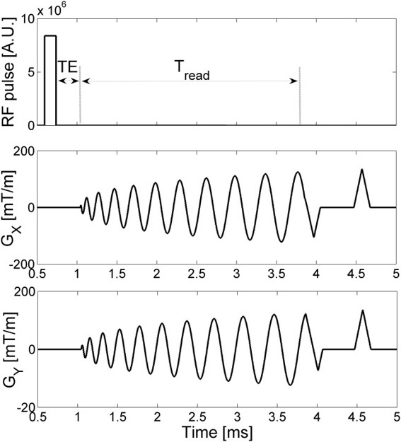
Purpose: To implement and optimize a single-shot spiral encoding strategy for rapid 2D IDEAL projection imaging of hyperpolarized (Hp) 129Xe in the gas phase, and in the pulmonary tissue (PT) and red blood cells (RBCs) compartments of the rat lung, respectively. Theory and Methods: A theoretical and experimental point spread function analysis was used to optimize the spiral kspace read-out time in a phantom. Hp 129Xe IDEAL images from five healthy rats were used to: (i) optimize flip angles by a Bloch equation analysis using measured kinetics of gas exchange and (ii) investigate the feasibility of the approach to characterize the exchange of Hp 129Xe.
Results: A read-out time equal to approximately 1.8 T 2 was found to
provide the best trade-off between spatial resolution and
signal-to-noise ratio (SNR). Spiral IDEAL approaches that use the
entire dissolved phase magnetization should give an SNR improvement
of a factor of approximately three compared with Cartesian
approaches with similar spatial resolution. The IDEAL strategy
allowed imaging of gas, PT, and RBC compartments with sufficient SNR
and temporal resolution to permit regional gas exchange measurements
in healthy rats.
Conclusion: Single-shot spiral IDEAL imaging of gas, PT and RBC
compartments and gas exchange is feasible in rat lung using Hp
129Xe. Magn Reson Med 76:566–576, 2016. VC 2015 Wiley Periodicals,
Inc.
...

Ozkan Doganay & Tahreema Matin & Mitchell Chen & Minsuok Kim & Anthony McIntyre & Daniel R. McGowan & Kevin M. Bradley & Thomas Povey & Fergus V. Gleeson
Purpose: To derive lobar ventilation in patients with chronic obstructive pulmonary disease (COPD) using a rapid time-series hyperpolarized xenon-129 (HPX) magnetic resonance imaging (MRI) technique and compare this to ventilation/perfusion singlephoton emission computed tomography (V/Q-SPECT), correlating the results with high-resolution computed tomography (CT) and pulmonary function tests (PFTs). Materials and methods Twelve COPD subjects (GOLD stages I–IV) participated in this study and underwent HPX-MRI, V/QSPECT/CT, high-resolution CT, and PFTs. HPX-MRI was performed using a novel time-series spiral k-space sampling approach. Relative percentage ventilations were calculated for individual lobe for comparison to the relative SPECT lobar ventilation and perfusion. The absolute HPX-MRI percentage ventilation in each lobe was compared to the absolute CT percentage emphysema score calculated using a signal threshold method. Pearson’s correlation and linear regression tests were performed to compare each imaging modality.
Results: Strong correlations were found between the relative lobar percentage ventilation with HPX-MRI and percentage ventilation SPECT (r = 0.644; p < 0.001) and percentage perfusion SPECT (r=0.767; p < 0.001). The absolute CT percentage emphysema and HPX percentage ventilation correlation was also statistically significant (r=0.695, p < 0.001). The whole lung HPX percentage ventilation correlated with the PFT measurements (FEV1 with r=− 0.886, p < 0.001*, and FEV1/FVC with r=− 0.861, p < 0.001*) better than the whole lung CT percentage emphysema score (FEV1 with r=− 0.635, p=0.027; and FEV1/FVC with r=− 0.652, p=0.021).
Conclusion:Lobar ventilation with HPX-MRI showed a strong correlation with lobar ventilation and perfusion measurements derived from SPECT/CT, and is better than the emphysema score obtained with high-resolution CT. ...
Ozkan Doganay, Tahreema N. Matin, Anthony Mcintyre, Brian Burns, Rolf F. Schulte, Fergus V. Gleeson, and Daniel Bulte
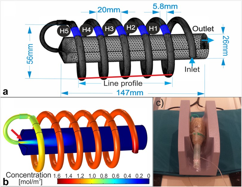
Purpose To develop and optimize a rapid dynamic hyperpolarized 129Xe ventilation (DXeV) MRI protocol and investigate the feasibility of capturing pulmonary signal-time curves in human lungs. Theory and Methods Spiral k-space trajectories were designed with the number of interleaves Nint = 1, 2, 4, and 8 corresponding to voxel sizes of 8 mm, 5 mm, 4 mm, and 2.5 mm, respectively, for field of view = 15 cm. DXeV images were acquired from a gas-flow phantom to investigate the ability of Nint = 1, 2, 4, and 8 to capture signal-time curves. A finite element model was constructed to investigate gas-flow dynamics corroborating the experimental signal-time curves. DXeV images were also carried out in six subjects (three healthy and three chronic obstructive pulmonary disease subjects).
Results: DXeV images and numerical modelling of signal-time curves permitted the quantification of temporal and spatial resolutions for different numbers of spiral interleaves. The two-interleaved spiral (Nint = 2) was found to be the most time-efficient to obtain DXeV images and signal-time curves of whole lungs with a temporal resolution of 624 ms for 13 slices. Signal-time curves were well matched in three healthy volunteers. The Spearman's correlations of chronic obstructive pulmonary disease subjects were statistically different from three healthy subjects (P < 0.05) ...
Conclusion The Nint = 2 spiral demonstrates the successful acquisition of DXeV images and signal-time curves in healthy subjects and chronic obstructive pulmonary disease patients. Magn Reson Med 79:2597–2606, 2018. © 2017 The Authors Magnetic Resonance in Medicine published by Wiley Periodicals, Inc. on behalf of International Society for Magnetic Resonance in Medicine. This is an open access article under the terms of the Creative Commons Attribution License, which permits use, distribution and reproduction in any medium, provided the original work is properly cited.
Ozkan Doganay, Elaine Stirrat, Charles McKenzie, Rolf F. Schulte, Giles E. Santyr
Purpose: To assess the feasibility of hyperpolarized (HP) 129Xe MRI for detection of early stage radiation-induced lung injury (RILI) in a rat model involving unilateral irradiation by assessing differences in gas exchange dynamics between irradiated and unirradiated lungs. Methods: The dynamics of gas exchange between alveolar air space and pulmonary tissue (PT), PT and red blood cells (RBCs) was measured using single-shot spiral iterative decomposition of water and fat with echo asymmetry and least-squares estimation images of the right and left lungs of two age-matched cohorts of Sprague Dawley rats. The first cohort (n = 5) received 18 Gy irradiation to the right lung using a 60Co source and the second cohort (n = 5) was not irradiated and served as the healthy control. Both groups were imaged two weeks following irradiation when radiation pneumonitis (RP) was expected to be present. The gas exchange data were fit to a theoretical gas exchange model to extract measurements of pulmonary tissue thickness (LPT) and relative blood volume (VRBC) from each of the right and left lungs of both cohorts. Following imaging, lung specimens were retrieved and percent tissue area (PTA) was assessed histologically to confirm RP and correlate with MRI measurements.
Results: Statistically significant differences in LPT and VRBC were observed between the irradiated and non-irradiated cohorts. In particular, LPT of the right and left lungs was increased approximately 8.2% and 5.0% respectively in the irradiated cohort. Additionally, VRBC of the right and left lungs was decreased approximately 36.1% and 11.7% respectively for the irradiated cohort compared to the non-irradiated cohort. PTA measurements in both right and left lungs were increased in the irradiated group compared to the non-irradiated cohort for both the left (P < 0.05) and right lungs (P < 0.01) confirming the presence of RP. PTA measurements also correlated with the MRI measurements for both the non-irradiated (r=0.79, P < 0.01) and irradiated groups (r=0.91, P < 0.01).
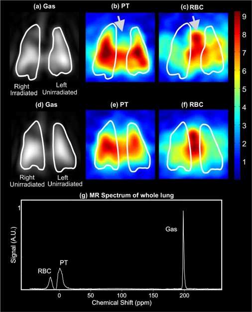
Conclusions: Regional RILI can be detected two weeks post-irradiation using HP 129Xe MRI and analysis of gas exchange curves. This approach correlates well with histology and can potentially be used clinically to assess radiation pneumonitis associated with early RILI to improve radiation therapy outcomes. Representative coronal gas, PT, and RBC images from an irradiated [(a)–(c)] and nonirradiated [(d)–(f)] animal obtained using three-point IDEAL for gas-PT and gas-RBC separately with a TR of 100 ms; (g) MR spectrum of rat lungs after inhaling HP 129Xe using a spectrally selective RF pulse that excites the gas phase with a low flip angle (18°) and the dissolved phases with a high flip angle (75°). The white contour lines in (a)–(f) represent the masks for the left and right lungs used for region-of-interest analysis. The strong signal in the center of the RBC image (c) as shown by the arrow appears to correspond to the left atrium, ventricle, and arch of the aorta.

Address:
Ege University Science and Technology Centre, 172/1, Bornova/İzmirPhone:
TR +90 531 741 03 94